


Bilevel PAP and APRV Ventilation in Critical Care: Principles, Clinical Applications, and Current Evidence
Clinical Technology Insights | Chenwei Medical ICU Ventilation Solutions
Introduction: Optimizing Lung Protection and Patient–Ventilator Synchrony in Modern Intensive Care
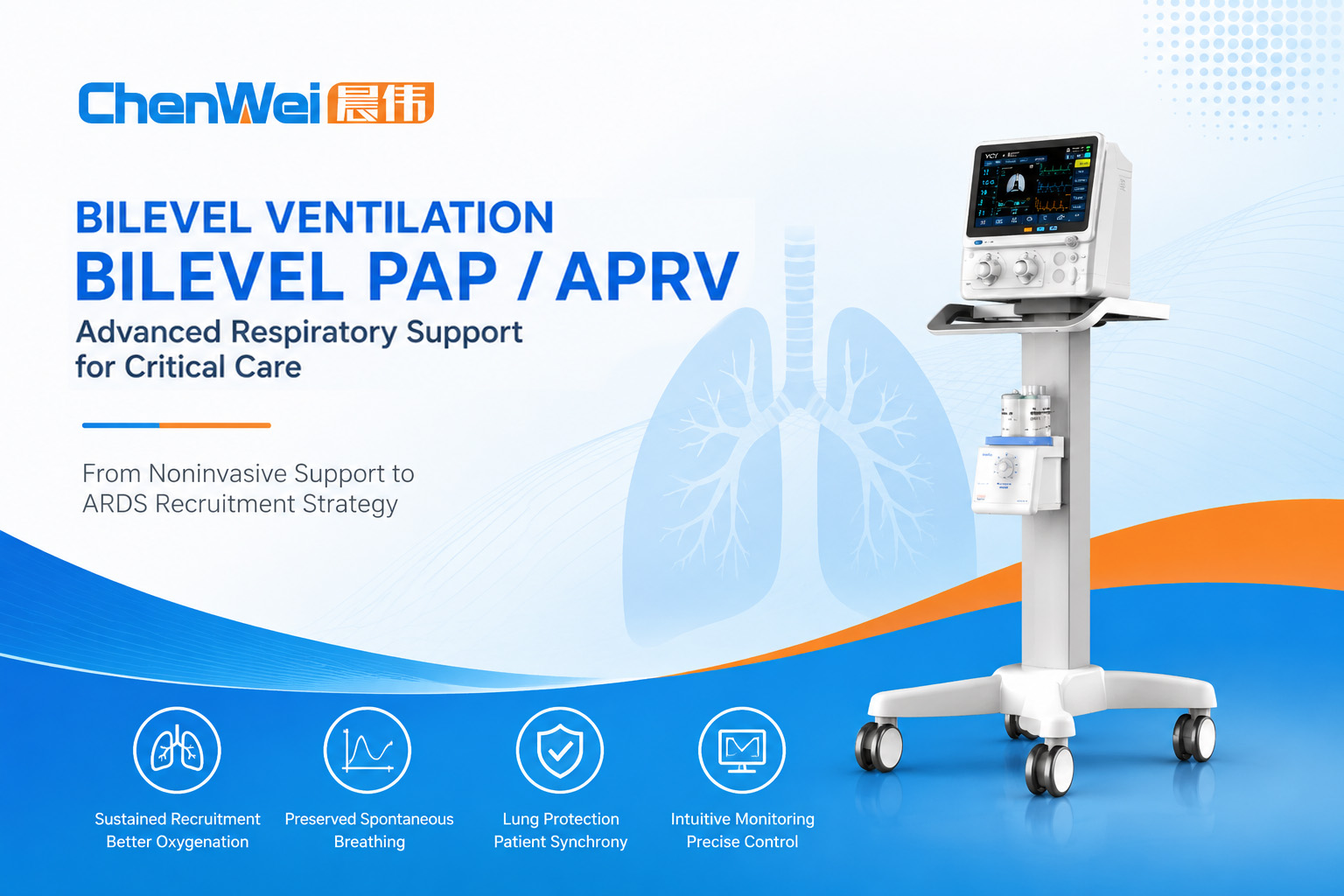
This article is intended for healthcare professionals and provides an overview of ventilation principles, clinical applications, and commonly reported parameter considerations. Ventilator settings should always be individualized according to patient condition, respiratory mechanics, institutional protocols, and clinical judgment.
Modern ICU ventilators must balance respiratory support with lung protection by providing precise pressure control, adaptive triggering, and advanced monitoring capabilities to meet the diverse needs of critically ill patients. As clinicians manage increasingly complex respiratory conditions—including acute exacerbations of chronic obstructive pulmonary disease (COPD), acute respiratory distress syndrome (ARDS), obesity hypoventilation syndrome (OHS), and other forms of acute respiratory failure—the challenge extends beyond simply improving oxygenation or carbon dioxide removal. Equally important is minimizing ventilator-induced lung injury (VILI), maintaining patient comfort, and promoting effective patient–ventilator synchrony.
To meet these clinical demands, modern ICU ventilators have evolved far beyond conventional volume-controlled and pressure-controlled ventilation. Advanced pressure-targeted ventilation strategies now enable greater flexibility by allowing spontaneous breathing while maintaining stable airway pressures. Among these, bilevel positive airway pressure (bilevel PAP) ventilation and airway pressure release ventilation (APRV) are widely used ventilation strategies for selected patient populations, each serving distinct clinical purposes depending on respiratory physiology and treatment goals.
Although different ventilator manufacturers may use proprietary names for bilevel ventilation modes, the underlying physiological principles are similar. These strategies utilize two preset airway pressure levels while permitting spontaneous breathing throughout most or all of the respiratory cycle, thereby improving alveolar recruitment, reducing the work of breathing, and potentially enhancing gas exchange.
Understanding the physiological rationale, parameter selection, waveform interpretation, and clinical indications of these ventilation strategies is essential for optimizing respiratory support in critically ill patients. This article reviews the principles of bilevel PAP ventilation and APRV while discussing practical parameter adjustment and clinical applications based on current evidence and modern ICU ventilator technology.
1. Understanding Bilevel Positive Airway Pressure Ventilation
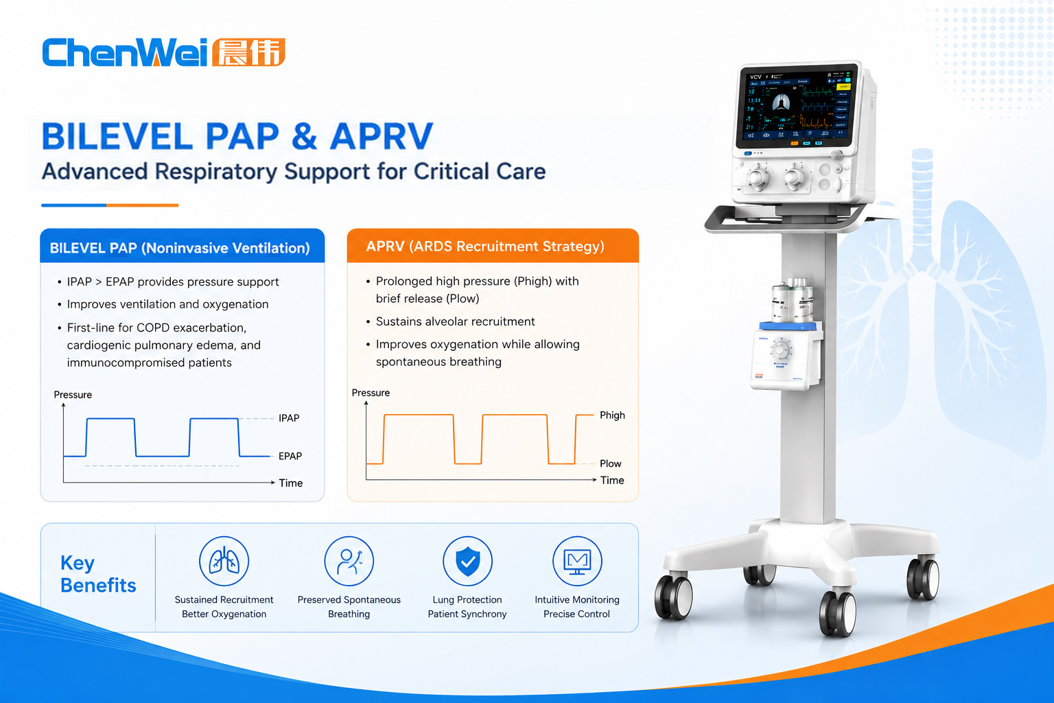
Bilevel positive airway pressure (bilevel PAP) ventilation is a pressure-targeted ventilation strategy that alternates between two preset airway pressure levels while allowing spontaneous breathing throughout the respiratory cycle.
Unlike conventional pressure support ventilation, which provides assistance only during spontaneous inspiration, bilevel ventilation maintains continuous positive airway pressure at two different pressure levels:
High airway pressure (PHigh) to promote alveolar recruitment and improve oxygenation
Low airway pressure (PLow) to facilitate exhalation and carbon dioxide elimination
Because spontaneous breathing is permitted at either pressure level, bilevel ventilation can improve patient comfort, preserve diaphragmatic activity, and enhance ventilation-perfusion matching.
1.1 Different Terminology Used by Ventilator Manufacturers
One source of confusion in clinical practice is that different ventilator manufacturers use different proprietary or platform-specific names for ventilation modes based on similar physiological principles.
Examples include:
Manufacturer-specific bilevel ventilation mode names
Biphasic positive airway pressure
BiLevel
DuoPAP
Other proprietary ventilation mode names
Despite differences in terminology, many of these modes are based on the same fundamental concept of alternating between two preset airway pressure levels while allowing spontaneous breathing.
Throughout this article, the generic term bilevel positive airway pressure (bilevel PAP) ventilation is used whenever discussing the underlying ventilation strategy rather than manufacturer-specific implementations.
1.2 Fundamental Physiological Principles
Regardless of nomenclature, bilevel ventilation is characterized by four primary parameters:
PHigh – upper airway pressure level
PLow – lower airway pressure level
THigh – duration spent at the high-pressure level
TLow – duration spent at the low-pressure level
Together, these parameters determine lung recruitment, ventilation efficiency, oxygenation, carbon dioxide clearance, and patient–ventilator synchrony.
Compared with conventional mandatory ventilation, bilevel ventilation offers several physiological advantages:
Continuous alveolar recruitment
Reduced cyclic alveolar collapse
Preservation of spontaneous breathing
Lower work of breathing
Improved ventilation–perfusion (V/Q) matching
Potential reduction in sedation requirements by allowing spontaneous breathing and improving patient comfort in appropriately selected patients
Although bilevel ventilation and APRV are both based on time-cycled switching between two preset airway pressure levels and share similar fundamental parameters (PHigh, PLow, THigh, and TLow), their clinical objectives and implementation strategies differ.
Conventional bilevel ventilation is generally used to provide pressure support, improve ventilation, and enhance patient comfort by allowing spontaneous breathing at both pressure levels. In contrast, APRV is primarily designed as an alveolar recruitment strategy that maintains a sustained high airway pressure for most of the respiratory cycle, with brief pressure releases to facilitate carbon dioxide elimination while preserving spontaneous breathing whenever clinically appropriate.
Therefore, APRV should not be considered simply as bilevel ventilation with a prolonged THigh. Instead, it represents a distinct ventilation strategy with a different physiological approach to lung recruitment, respiratory mechanics, and patient management.
1.3 Relationship Between NIV Bilevel PAP and ICU Bilevel Invasive Ventilation
Although the terms “bilevel PAP” and “bilevel ventilation” are often used interchangeably in clinical discussions, they may refer to different applications depending on the ventilation setting.
In noninvasive ventilation (NIV), bilevel PAP generally refers to pressure support delivered through a patient interface such as an oronasal or nasal mask. The commonly used parameters are inspiratory positive airway pressure (IPAP) and expiratory positive airway pressure (EPAP). The difference between IPAP and EPAP determines the level of pressure support and primarily influences tidal volume and carbon dioxide elimination.
In invasive mechanical ventilation using ICU ventilators, bilevel ventilation modes are typically described using parameters such as high airway pressure (PHigh), low airway pressure (PLow), high-pressure duration (THigh), and low-pressure duration (TLow). These modes allow spontaneous breathing while alternating between two controlled pressure levels delivered through an artificial airway.
Although both approaches utilize two pressure levels, their clinical goals are different. NIV bilevel PAP is primarily used to provide ventilatory assistance and reduce the work of breathing without intubation, whereas invasive bilevel ventilation is designed for critically ill patients requiring advanced respiratory support, including those with complex lung mechanics and severe respiratory failure.
Understanding this distinction is important because similar terminology may represent different ventilator functions depending on the clinical environment and ventilator platform.
2. Noninvasive Bilevel PAP Ventilation: Clinical Applications and Initial Settings
Noninvasive ventilation (NIV using bilevel PAP) has become a standard treatment strategy for acute hypercapnic respiratory failure and selected cases of acute hypoxemic respiratory failure.
Instead of delivering ventilation through an artificial airway, NIV provides pressure support through a well-fitted oronasal or nasal interface while preserving normal airway defense mechanisms.
In this setting:
Inspiratory Positive Airway Pressure (IPAP) provides inspiratory assistance.
Expiratory Positive Airway Pressure (EPAP) maintains end-expiratory lung volume and prevents alveolar collapse.
Pressure Support (PS) equals the difference between IPAP and EPAP.
Mathematically:
Pressure Support = IPAP − EPAP
Higher pressure support generally increases tidal volume and carbon dioxide elimination, whereas EPAP primarily improves oxygenation by increasing functional residual capacity (FRC) and maintaining alveolar recruitment.
2.1 Commonly Used Initial Parameter Ranges
The following values represent commonly reported clinical starting ranges rather than universal recommendations. Initial settings should always be individualized according to respiratory mechanics, blood gas analysis, patient comfort, and institutional protocols.
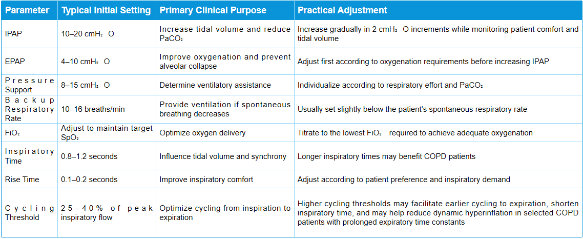
Rather than following fixed numerical values, clinicians should individualize ventilator settings according to respiratory mechanics, blood gas analysis, waveform assessment, and overall patient response.
2.2 Major Clinical Indications
Current international guidelines support bilevel PAP ventilation as an effective noninvasive respiratory support strategy in appropriately selected patients.
Acute Exacerbation of COPD
Acute hypercapnic respiratory failure caused by COPD remains the strongest evidence-based indication for noninvasive bilevel PAP ventilation.
Large randomized controlled trials and international guidelines have consistently shown that appropriately applied noninvasive ventilation can reduce the need for endotracheal intubation, shorten hospital stay, and improve clinical outcomes in selected patients with acute hypercapnic respiratory failure secondary to COPD.
Acute Cardiogenic Pulmonary Edema
Patients with cardiogenic pulmonary edema benefit from positive airway pressure through improved alveolar recruitment, reduced preload and afterload, and decreased work of breathing.
Compared with continuous positive airway pressure (CPAP) alone, bilevel PAP ventilation may provide additional ventilatory assistance in patients with concurrent hypercapnia or significant respiratory muscle fatigue.
Immunocompromised Patients
For selected immunocompromised patients experiencing acute respiratory failure, early application of noninvasive respiratory support may reduce the need for endotracheal intubation and its associated infectious complications, provided that patients are closely monitored and appropriate escalation criteria are followed.
Postoperative Respiratory Failure
Following major thoracic or abdominal surgery, carefully selected patients with postoperative respiratory insufficiency may benefit from bilevel PAP ventilation to improve lung expansion, reduce atelectasis, and decrease the likelihood of reintubation.
However, careful patient selection and continuous reassessment remain essential to avoid delaying invasive mechanical ventilation when clinically indicated.
3. Airway Pressure Release Ventilation (APRV): An Advanced Lung Recruitment Strategy
Airway Pressure Release Ventilation (APRV) is an advanced pressure-targeted ventilation strategy designed to maximize alveolar recruitment while preserving spontaneous breathing whenever possible.
Unlike conventional mechanical ventilation, APRV maintains the lungs at a sustained high airway pressure for most of the respiratory cycle, with only brief pressure releases to facilitate carbon dioxide elimination. This approach aims to improve oxygenation, reduce cyclic alveolar collapse, and support lung-protective ventilation in selected patients with moderate-to-severe acute respiratory distress syndrome (ARDS).
When spontaneous breathing is preserved, APRV may also improve diaphragmatic function, enhance ventilation–perfusion matching, and reduce the need for deep sedation.
Although APRV has shown physiological advantages in many studies, current international guidelines generally regard it as an alternative or rescue ventilation strategy rather than a replacement for conventional lung-protective ventilation.
3.1 Physiological Principles of APRV
APRV operates using the same four fundamental parameters as bilevel ventilation:
PHigh – high airway pressure level
PLow – low airway pressure level
THigh – duration spent at high pressure
TLow – duration spent at low pressure
Although APRV shares several technical characteristics with bilevel pressure ventilation, its therapeutic philosophy differs. Rather than simply alternating between two pressure levels, APRV aims to maximize alveolar recruitment by maintaining the lung at a relatively high continuous pressure for most of the respiratory cycle, while allowing only brief pressure releases to facilitate carbon dioxide elimination. Whenever clinically appropriate, spontaneous breathing is encouraged throughout both pressure phases to support respiratory muscle activity and improve patient–ventilator interaction.
This creates three important physiological effects:
Sustained Alveolar Recruitment
Maintaining the lungs at an elevated pressure for several seconds allows unstable alveoli to remain open, thereby improving oxygenation while minimizing repetitive opening and closing of alveolar units.
Brief pressure release to facilitate ventilation and CO₂ clearance
A short release to PLow allows partial exhalation and carbon dioxide removal without permitting complete lung collapse.
This concept distinguishes APRV from traditional mandatory ventilation, where expiration occupies a much larger proportion of the respiratory cycle.
Preservation of Spontaneous Breathing
Whenever clinically appropriate, spontaneous breathing is encouraged throughout both pressure phases.
Maintaining spontaneous breathing, when clinically appropriate, may provide several physiological benefits:
Improve ventilation–perfusion distribution
Support diaphragmatic activity and reduce disuse-related respiratory muscle weakness
Facilitate patient–ventilator interaction
Potentially reduce the need for deep sedation in selected patients
The effects of spontaneous breathing on cardiovascular function may vary depending on intrathoracic pressure, lung mechanics, and overall hemodynamic status.
3.2 Commonly Reported Initial Parameter Ranges
The following ranges represent commonly reported clinical starting points rather than fixed recommendations.
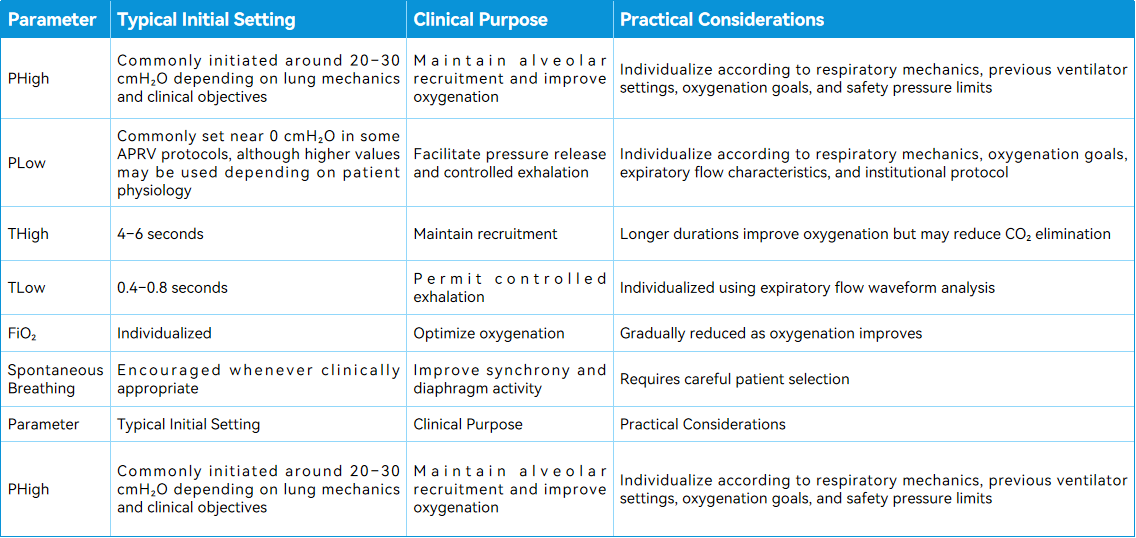
In APRV, PHigh selection should not be determined by a single numerical target. Clinicians should consider oxygenation response, respiratory mechanics, chest wall compliance, spontaneous tidal volume, and potential risks associated with excessive airway pressure.
It should be noted that APRV parameter selection varies among institutions, ventilator platforms, and clinician experience. For example, some published APRV protocols initiate PLow at or near 0 cmH₂O with a short release phase, whereas other clinicians may select a higher PLow based on respiratory mechanics, oxygenation requirements, expiratory flow characteristics, and institutional practice. Therefore, PLow should be considered a patient-specific parameter rather than a universal fixed value.
Ventilator settings should always be individualized according to respiratory mechanics, gas exchange, hemodynamic status, and continuous waveform assessment.
3.3 Fine-Tuning TLow: The Most Critical APRV Adjustment
Among all APRV parameters, TLow is generally considered the most important for achieving effective lung protection.
If TLow is excessively long, excessive lung volume may be lost during exhalation, leading to alveolar derecruitment.
Conversely, if TLow is too short, inadequate carbon dioxide elimination may result in progressive hypercapnia.
Rather than relying solely on preset numerical values, many experienced clinicians individualize TLow by assessing the expiratory flow waveform. A commonly described approach is to terminate the release phase when expiratory flow has decelerated to approximately 50–75% of peak expiratory flow, although the optimal target may vary according to respiratory mechanics, disease severity, spontaneous breathing activity, and institutional practice.
Waveform Interpretation
Appropriate TLow
Expiratory flow is interrupted before complete lung emptying
Maintenance of end-expiratory lung volume
Stable oxygenation and acceptable CO₂ clearance
No evidence of significant air trapping
Excessively Long TLow
Clinical findings may include:
Complete expiratory flow reaching baseline before pressure release ends
Loss of end-expiratory lung volume
Increased alveolar derecruitment
Potential deterioration in oxygenation
Excessively Short TLow
Potential consequences include:
Reduced CO₂ clearance due to insufficient release volume
Progressive hypercapnia
Increased airway pressure due to incomplete pressure release
Possible patient discomfort or dyssynchrony
Accordingly, waveform interpretation should guide parameter adjustment rather than relying solely on preset time values.
3.4 Practical APRV Weaning Strategy
Successful liberation from APRV typically involves gradual reduction of ventilatory support while preserving spontaneous breathing.
Several weaning strategies have been described in the literature. One commonly adopted approach includes the following four stages, although institutional protocols may differ.
Step 1 – Stabilization
Maintain adequate oxygenation using relatively high PHigh with prolonged THigh while ensuring patient comfort and acceptable gas exchange.
Step 2 – Gradual Reduction of PHigh
As oxygenation improves, decrease PHigh gradually, commonly by approximately 2 cmH₂O at a time, while maintaining adequate lung recruitment.
Step 3 – Progressive Extension of THigh
Increasing THigh reduces the frequency of pressure releases, allowing patients to assume a greater proportion of spontaneous ventilation.
Step 4 – Transition to Conventional Assisted Ventilation
Once airway pressures and oxygen requirements have sufficiently decreased, patients may be transitioned to pressure support ventilation (PSV) or another spontaneous breathing mode before completing a spontaneous breathing trial (SBT) according to institutional protocols.
4.Comparing Bilevel PAP Ventilation, APRV, and Conventional Lung-Protective Ventilation
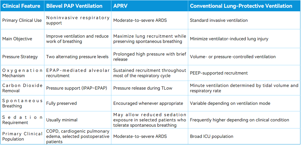
Current Evidence
Although APRV provides several physiological advantages—including sustained alveolar recruitment, preservation of spontaneous breathing, and potential improvement in patient–ventilator interaction—the current clinical evidence remains insufficient to establish clear superiority over conventional lung-protective ventilation strategies for major patient-centered outcomes.
Clinical studies and meta-analyses have reported potential benefits in areas such as oxygenation, respiratory mechanics, and sedation requirements; however, differences in APRV protocols, patient selection, clinician experience, and study methodology have limited definitive conclusions regarding mortality reduction or consistent improvement in long-term outcomes.
Consequently, contemporary international guidelines continue to recommend low tidal volume ventilation as the standard first-line strategy for most patients with ARDS, based on established evidence demonstrating reduced mortality compared with traditional tidal volume approaches. APRV may be considered as an alternative strategy in selected patients, particularly in experienced centers with appropriate monitoring capabilities.
5. Optimizing Patient–Ventilator Synchrony During Bilevel Ventilation
Patient–ventilator synchrony plays a critical role in determining the effectiveness of mechanical ventilation. Poor synchrony can increase the work of breathing, compromise patient comfort, prolong mechanical ventilation, and potentially increase the need for sedation.
Modern ICU ventilators employ advanced triggering algorithms, leak compensation technologies, and waveform monitoring tools to help clinicians identify and correct synchrony issues in real time.
5.1 Common Synchrony Challenges During Noninvasive Bilevel PAP Ventilation
Mask Leak and Delayed Cycling
Mask leakage remains one of the most common causes of ineffective noninvasive ventilation.
Excessive leakage may interfere with inspiratory flow detection, delaying the transition from inspiration to expiration. As a result, patients may attempt to exhale while inspiratory pressure is still being delivered, producing discomfort and poor synchrony.
Recommended interventions include:
Improving mask fit and interface selection
Adjusting cycling sensitivity
Utilizing ventilator leak-compensation functions where available
Auto-Triggering
Large leaks, excessive circuit movement, or water condensation may generate false trigger signals, causing unnecessary ventilator breaths.
Potential corrective measures include:
Increasing trigger sensitivity thresholds appropriately
Removing excess condensate from the breathing circuit
Inspecting tubing and patient interfaces for leakage
Excessive Pressure Support
Excessive pressure support may deliver tidal volumes greater than clinically necessary, increasing patient discomfort and reducing spontaneous respiratory effort.
Management typically involves gradual reduction of pressure support while continuously assessing tidal volume, respiratory rate, patient comfort, and arterial blood gases.
5.2 Synchrony Considerations During APRV
Because spontaneous breathing is encouraged during APRV, careful waveform assessment remains essential.
Incomplete Exhalation
An excessively short TLow may prevent adequate exhalation, resulting in progressive air trapping and intrinsic positive end-expiratory pressure (auto-PEEP).
When this occurs, clinicians should reassess:
TLow duration
PHigh level
Respiratory mechanics
Expiratory flow waveform
Excessive Respiratory Drive
Patients with severe respiratory distress may exhibit vigorous inspiratory or expiratory efforts despite ventilator assistance.
Persistent dyssynchrony should prompt reassessment of:
Sedation and analgesia
Ventilator parameter settings
Underlying disease progression
Patient comfort and respiratory drive
Optimizing synchrony requires integrating clinical assessment with continuous waveform interpretation rather than relying solely on numerical ventilator parameters.
6. Clinical Applications of Bilevel Ventilation
6.1 Acute Exacerbation of COPD
Acute exacerbation of COPD complicated by hypercapnic respiratory failure remains the strongest evidence-based indication for noninvasive bilevel PAP ventilation.
Early initiation in appropriately selected patients has consistently demonstrated reductions in:
Need for endotracheal intubation
Treatment failure requiring escalation of respiratory support
Hospital mortality in selected patient populations
Complications associated with invasive mechanical ventilation
Initial NIV settings should be individualized according to the severity of respiratory failure, degree of hypercapnia, patient tolerance, respiratory mechanics, and clinical response.
Typical initial settings may include:
IPAP: 12–16 cmH₂O
EPAP: 4–6 cmH₂O
Backup respiratory rate: 10–12 breaths/min
Supplemental oxygen should be titrated carefully to achieve the recommended oxygen saturation target range while avoiding unnecessary excessive oxygen exposure, particularly in patients with chronic hypercapnic respiratory failure.
6.2 Obesity Hypoventilation Syndrome (OHS)
Patients with obesity hypoventilation syndrome frequently present with concomitant obstructive sleep apnea and chronic hypercapnic respiratory failure.
Bilevel PAP ventilation may improve:
Alveolar ventilation
Carbon dioxide elimination
Sleep quality
Daytime symptoms
Treatment parameters should be individualized according to respiratory mechanics, gas exchange, and sleep study findings when available.
6.3 Neuromuscular Disorders
Patients with neuromuscular disorders such as amyotrophic lateral sclerosis (ALS), spinal muscular atrophy (SMA), or muscular dystrophy may benefit from long-term noninvasive ventilatory support.
In these patients, bilevel PAP ventilation aims to:
Reduce respiratory muscle workload
Improve nocturnal ventilation
Enhance quality of life
Delay progression to invasive ventilation when clinically appropriate
Treatment goals should always be individualized according to disease stage, patient preference, and multidisciplinary clinical assessment.
7. Conclusion
Successful mechanical ventilation extends beyond selecting an appropriate ventilation mode. Achieving optimal clinical outcomes requires careful integration of respiratory physiology, individualized parameter adjustment, continuous waveform interpretation, and ongoing reassessment of patient response.
Bilevel positive airway pressure ventilation provides an effective noninvasive respiratory support strategy for carefully selected patients with acute respiratory failure.
APRV offers an alternative lung recruitment approach for selected patients with moderate-to-severe ARDS when managed by experienced critical care teams. Careful monitoring of spontaneous tidal volume, respiratory effort, respiratory mechanics, and patient–ventilator interaction remains essential.
Modern ICU ventilators equipped with precise pressure control, responsive triggering systems, comprehensive waveform monitoring, and advanced ventilation modes enable clinicians to individualize respiratory support across a wide spectrum of critical care scenarios.
Chenwei Medical provides ICU ventilation solutions integrating reliable respiratory technology, intuitive operation, and advanced monitoring capabilities to support individualized respiratory care. By integrating engineering innovation with evidence-informed respiratory care principles, our goal is to support healthcare professionals in delivering safe and individualized ventilation strategies across diverse critical care environments.
Medical Disclaimer
This article is provided for educational and informational purposes only. It does not constitute medical advice and should not replace professional clinical judgment, institutional protocols, or evidence-based guidelines. Ventilator settings should always be individualized according to each patient's condition, respiratory mechanics, and the assessment of qualified healthcare professionals.
Clinical Practice Notice
The parameter ranges and clinical examples presented in this article represent commonly reported practices in the published literature and should not be interpreted as universal recommendations. Actual ventilator settings should always be determined by qualified healthcare professionals based on individual patient characteristics and current clinical guidelines.
Trademark Notice
Certain ventilation mode names referenced in this article may be registered trademarks or proprietary designations of their respective owners.
For example, BiPAP® is a registered trademark of Koninklijke Philips N.V. (or its affiliated companies). Other ventilation mode names mentioned in this article may also be registered trademarks or proprietary designations of their respective owners.
These names are referenced solely for the purpose of explaining commonly used clinical terminology. Their inclusion does not imply any affiliation with, endorsement by, sponsorship from, or association with the respective trademark owners. Unless specifically referring to manufacturer-defined terminology, this article uses the generic term "bilevel positive airway pressure (bilevel PAP) ventilation."
References
Rochwerg B, Brochard L, Elliott MW, Hess D, Hill NS, Nava S, et al. Official ERS/ATS clinical practice guidelines: noninvasive ventilation for acute respiratory failure. Eur Respir J. 2017;50(2):1602426.
Lightowler JV, Wedzicha JA, Elliott MW, Ram FSF. Non-invasive positive pressure ventilation to treat respiratory failure resulting from exacerbations of chronic obstructive pulmonary disease. Cochrane Database Syst Rev. 2003;(1):CD004104.
Gray A, Goodacre S, Newby DE, Masson M, Sampson F, Nicholl J; 3CPO Trialists. Noninvasive ventilation in acute cardiogenic pulmonary edema. N Engl J Med. 2008;359(2):142–151.
Hilbert G, Gruson D, Vargas F, Valentino R, Gbikpi-Benissan G, Dupon M, et al. Noninvasive ventilation in immunosuppressed patients with pulmonary infiltrates, fever, and acute respiratory failure. N Engl J Med. 2001;344(7):481–487.
Jaber S, Lescot T, Futier E, Paugam-Burtz C, Seguin P, Ferrandière M, et al. Effect of noninvasive ventilation on tracheal reintubation among patients with hypoxemic respiratory failure following abdominal surgery: a randomized clinical trial. JAMA. 2015;313(23):2331–2339.
Putensen C, Mutz NJ, Putensen-Himmer G, Zinserling J. Spontaneous breathing during ventilatory support improves ventilation-perfusion distributions in patients with acute respiratory distress syndrome. Am J Respir Crit Care Med. 1999;159(4 Pt 1):1241–1248.
Fan E, Del Sorbo L, Goligher EC, Hodgson CL, Munshi L, Walkey AJ, et al. An Official American Thoracic Society/European Society of Intensive Care Medicine/Society of Critical Care Medicine Clinical Practice Guideline: Mechanical ventilation in adult patients with acute respiratory distress syndrome. Am J Respir Crit Care Med. 2017;195(9):1253–1263.
Global Initiative for Chronic Obstructive Lung Disease (GOLD). Global Strategy for Prevention, Diagnosis and Management of COPD: 2025 Report. Global Initiative for Chronic Obstructive Lung Disease; 2025.
Habashi NM. Other approaches to open-lung ventilation: airway pressure release ventilation. Crit Care Med. 2005;33(3 Suppl):S228–S240.
Mireles-Cabodevila E, Kacmarek RM. Should airway pressure release ventilation be the primary mode in ARDS? Respir Care. 2016;61(6):761–773.
Brower RG, Matthay MA, Morris A, Schoenfeld D, Thompson BT, Wheeler A; ARDS Network. Ventilation with lower tidal volumes as compared with traditional tidal volumes for acute lung injury and the acute respiratory distress syndrome. N Engl J Med. 2000;342(18):1301–1308.
Zhou Y, Jin X, Lv Y, et al. Early application of airway pressure release ventilation may reduce the duration of mechanical ventilation in acute respiratory distress syndrome. Intensive Care Medicine. 2017;43:1648–1657.
Carsetti A, Damiani E, Domizi R, et al. Airway pressure release ventilation during acute respiratory failure: a systematic review and meta-analysis. Critical Care. 2019;23:22.
Disclaimer: This article is intended solely for the exchange of professional knowledge and informational reference and does not constitute medical advice, diagnosis, or treatment recommendations, nor should it be construed as a guarantee or representation regarding the clinical efficacy or safety of any product. The academic perspectives, research data, and conclusions presented herein are based on the publicly available literature cited in the article. If any content herein involves or potentially infringes upon the legitimate rights or interests of any third party, please contact us. We will promptly review the matter and take appropriate action where warranted.
Related News