


Pressure-time, flow-time, and volume-time clues for a structured bedside review
Ventilator waveforms provide important clues about timing and patient-ventilator interaction, but they should not be interpreted as stand-alone diagnoses. Patient-ventilator synchrony means that patient effort, flow demand, and neural inspiratory timing are reasonably matched by ventilator triggering, gas delivery, and cycling.
Because no waveform pattern is normal across every patient and ventilation mode, waveform assessment should be combined with clinical evaluation. Review pressure-time, flow-time, and volume-time waveforms together when appropriate, then determine whether the mismatch is primarily related to triggering, flow delivery, cycling, or expiration.
Start with the Patient, Then the Graphics
A waveform should never outrank the patient. Assess oxygenation, ventilation, respiratory distress, chest and abdominal movement, level of consciousness, hemodynamics, and the airway and circuit.
The 2024 AARC Clinical Practice Guideline places patient-ventilator assessment within a broader patient-centered evaluation that includes physiology, physical examination, the artificial airway, respiratory mechanics, ventilator settings, and alarms [1]. Condensation, leaks, secretions, coughing, movement, and other circuit or patient factors can distort a trace and may mimic patient-ventilator asynchrony.
Read Pressure and Flow Together
Pressure-time waveforms can help identify trigger effort, inspiratory pressure distortion, and changes around cycling. Flow-time waveforms can show whether an inspiratory effort is followed by a supported breath, whether delivered flow appears adequate for patient demand, how closely consecutive machine breaths occur, and whether expiratory flow approaches baseline before the next breath.
Volume-time waveforms can help confirm volume trends and support assessment of leaks or incomplete expiration, although they rarely classify the patient-ventilator interaction on their own.
When learning to read ventilator waveforms, begin with pressure and flow displayed on the same time scale. Add volume when it helps answer a specific clinical question. A prospective study found relatively low sensitivity for detecting patient-ventilator asynchrony overall: report-level assessment outperformed breath-by-breath analysis (55% vs 22%), while experts outperformed nonexperts within breath-by-breath analysis (28% vs 16%) [2].
Review several consecutive breaths rather than relying on a single waveform.

What Closer Synchrony Looks Like
In a more synchronous sequence, patient effort is followed promptly by ventilator assistance, pressure and flow follow the expected pattern for the selected mode, and cycling occurs near the end of inspiratory effort.
Expiratory flow generally moves toward baseline before the next breath. These findings should be confirmed across several breaths and considered together with the patient's clinical condition. A smooth waveform alone does not prove adequate support, appropriate ventilation, or safe settings.

Figure 1. Illustrative teaching baseline for closer patient-ventilator synchrony.
Four Waveform Patterns Worth Recognizing
For each suspected pattern, compare the observed breath with the expected pressure, flow, and volume sequence for the selected mode. First identify where the expected sequence changes; then consider potential mechanisms and appropriate clinical responses.
1. Ineffective Triggering
Waveform Clue
Start with expiration. During an effective trigger, a small pressure dip or expiratory-flow deflection is followed by a pressure rise and positive inspiratory flow.
With ineffective triggering, a pressure or flow deflection may appear without a subsequent supported breath. Pressure and flow remain in expiration, suggesting that a patient inspiratory effort did not trigger the ventilator. Repeated ineffective efforts may result in a displayed respiratory rate that is lower than the patient's actual effort rate.
Clinical and Technical Assessment
Assess expiratory flow, intrinsic PEEP, trigger sensitivity, respiratory rate, tidal volume, minute ventilation, and gas exchange. After checking the circuit and airway, consider insufficient expiratory time, increased resistance, weak inspiratory effort, insensitive triggering, intrinsic PEEP, or excessive ventilatory assistance.
Chenwei Bedside Approach
Where supported by the specific ventilator configuration, waveform freeze and review functions can help clinicians examine brief pressure or flow deflections that may otherwise be difficult to see in real time. Trigger sensitivity can then be assessed and adjusted according to the patient's clinical condition and the ventilator mode.

Figure 2. Illustrative ineffective-triggering pattern showing an expiratory pressure or flow deflection without a subsequent supported breath.
2. Flow Insufficiency During Mechanical Ventilation
Waveform Clue
Read the pressure waveform during inspiration. With adequate flow, airway pressure follows the expected contour for the selected mode and flow pattern.
An inward pressure deflection or sustained scoop during inspiration may suggest that delivered flow is insufficient for patient demand, particularly when the pattern recurs across breaths and is consistent with the patient's clinical presentation.
Clinical and Technical Assessment
Confirm the pattern across multiple breaths, then assess respiratory drive, work of breathing, P0.1 when available, tidal and minute ventilation, gas exchange, and relevant pressure measurements such as peak, plateau, and driving pressure [3,7].
Consider potential contributors such as pain, anxiety, fever, acidosis, or a mismatch involving inspiratory flow, rise time, inspiratory time, or ventilation mode.
Dual-control modes with adaptive targets, such as PRVC or Autoflow, may adjust pressure delivery according to breath-by-breath conditions and should not be interpreted simply as conventional pressure-controlled ventilation.
Chenwei Bedside Approach
When available on the specific device, waveform review and adjustable inspiratory parameters can help clinicians evaluate whether the selected flow, rise time, or mode is appropriate for the patient's demand.
Chenwei T Series ventilators are designed to provide responsive flow and pressure delivery. Such features may support clinicians in assessing and optimizing patient-ventilator interaction, subject to the specific device configuration, operating instructions, and clinical context.

Figure 3. Illustrative pressure-time waveform in which an inward inspiratory deflection may suggest insufficient flow in a constant-flow volume-control example.
3. Double Triggering vs. Reverse Triggering
Waveform Clue and Clinical Distinction
Read the transition between consecutive breaths.
In a more synchronous sequence, one inspiratory phase is followed by clear expiration before the next inspiration. With double triggering, two consecutive assisted breaths may occur with only a short expiratory interval. Expiratory volume may not return fully toward baseline, and delivered volume can accumulate across the two breaths.
Double Triggering — Patient-Related Drive
Double triggering is often associated with high or prolonged patient inspiratory effort that outlasts the ventilator's inspiratory time, resulting in two consecutive machine-supported breaths and possible breath stacking [6].
Reverse Triggering — Ventilator-Related Timing
Reverse triggering refers to a ventilator-initiated breath followed by a patient diaphragmatic or respiratory muscle contraction. This phenomenon may involve ventilator-induced respiratory entrainment and requires distinction from patient-driven double triggering because the underlying mechanism and potential management strategies differ.
Clinical and Technical Assessment
Assess stacked exhaled volume, tidal volume relative to predicted body weight, peak, plateau, and driving pressures, as well as minute ventilation and the set or delivered respiratory rate [4].
Consider respiratory drive, premature cycling, inspiratory time, flow, and cycle-off criteria before adjusting ventilatory support.
When reverse triggering is suspected and the mechanism remains uncertain, additional monitoring such as esophageal pressure or electrical activity of the diaphragm may help characterize the interaction.
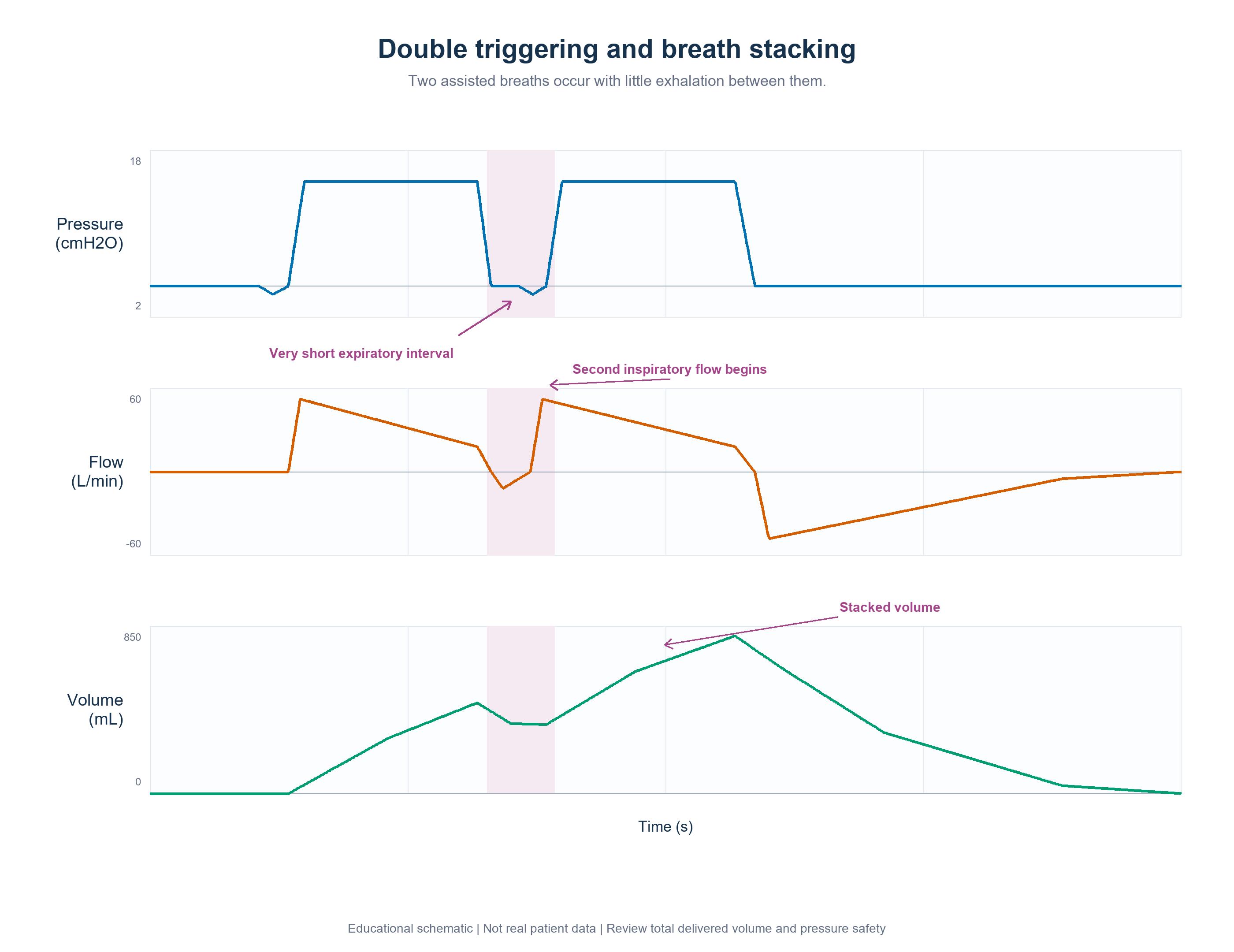
Figure 4. Illustrative double-triggering pattern showing two assisted breaths separated by a short expiratory interval and potential volume stacking.
Chenwei Bedside Approach
Where supported by the device, waveform freeze and review functions can help clinicians examine the interval between consecutive breaths and assess changes in delivered volume.
Adjustments to inspiratory time, cycling criteria, or ventilation mode should be based on the patient's respiratory effort, gas exchange, mechanics, and the specific ventilator's operating instructions.
4. Premature Cycling
Waveform Clue
Read the transition from inspiration to expiration.
With closer synchrony, ventilator cycling occurs near the end of patient inspiratory effort and expiratory flow proceeds smoothly.
With premature cycling, mechanical inspiration may end while patient inspiratory effort continues. A pressure deflection immediately after cycling, an abnormal early-expiratory flow pattern, or a rapid subsequent trigger may suggest that mechanical inspiratory time is shorter than neural inspiratory time.
Clinical and Technical Assessment
Assess inspiratory time, I:E ratio, cycle-off criteria, repeat triggering, respiratory rate, tidal and minute ventilation, gas exchange, and expiratory flow.
In flow-cycled pressure-support ventilation, the cycle-off threshold can be adjusted to influence mechanical inspiratory time. In time-cycled ventilation, inspiratory time may be adjusted when clinically appropriate [8].
The appropriate adjustment depends on the patient's respiratory mechanics, inspiratory effort, ventilation mode, and clinical condition.
Chenwei Bedside Approach
Where available, waveform freeze and comparison functions can assist clinicians in reviewing pressure and flow changes around cycling.
Cycle-off and inspiratory-time adjustments should be individualized rather than applied as fixed numerical recommendations. Any change should be evaluated by comparing the subsequent waveform with the patient's clinical response.
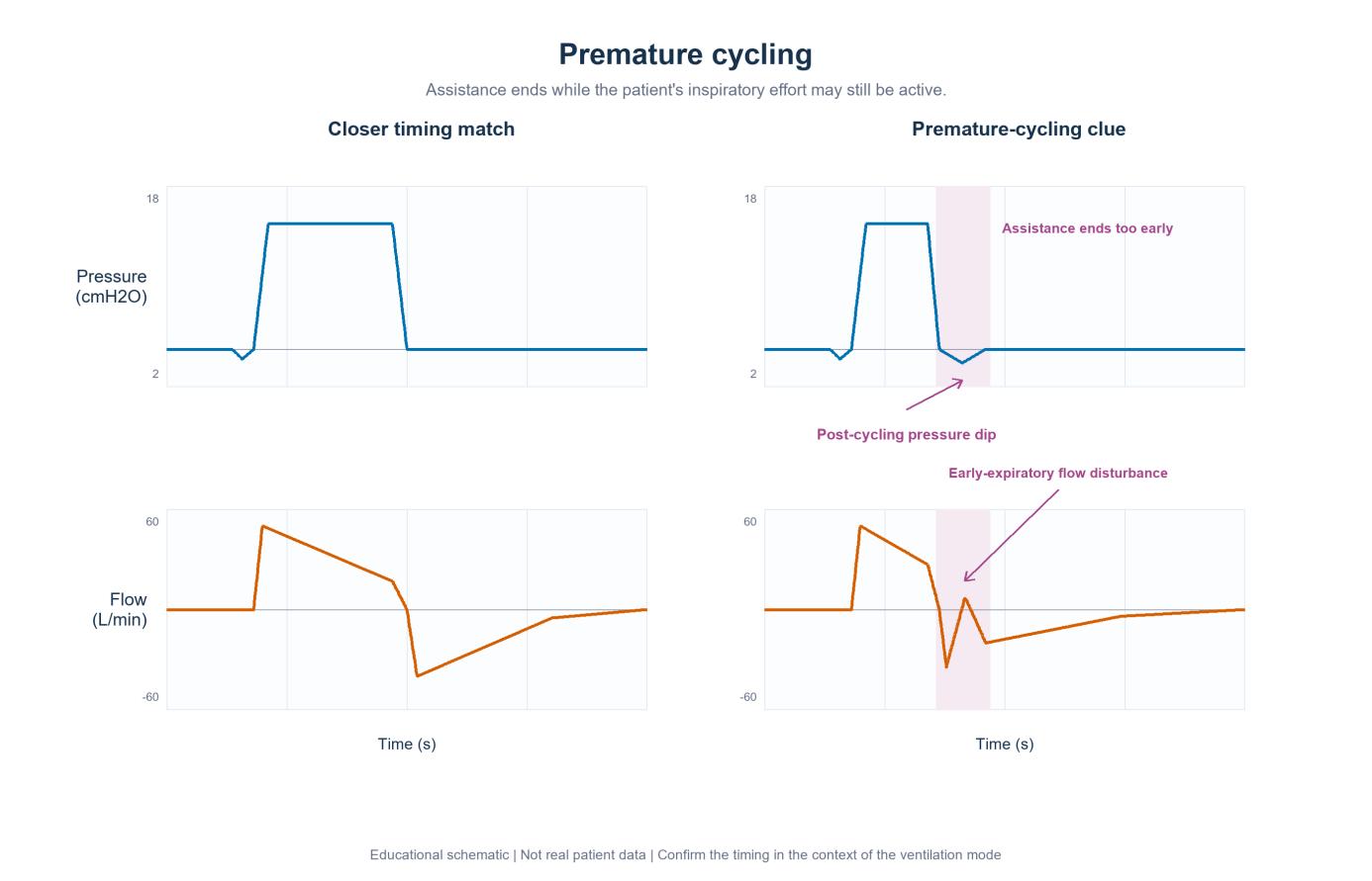
Figure 5. Illustrative premature-cycling pattern in which post-cycling pressure and flow disturbances may suggest continuing inspiratory effort.
Chenwei T Series: Waveform Review and Synchrony-Oriented Features
Chenwei ventilators incorporate waveform monitoring and adjustable ventilation parameters intended to support bedside assessment of patient-ventilator interaction.
Depending on the specific model and configuration, relevant functions may include:

These functions should be understood as tools for waveform assessment and ventilator adjustment rather than as independent methods for diagnosing or resolving patient-ventilator asynchrony.
Specific functions, parameter ranges, and performance characteristics should always be verified against the operating instructions and specifications for the individual Chenwei ventilator model.
Know What Standard Waveforms Cannot Settle
The 2026 SYNAPsE modified Delphi study identified several patient-ventilator asynchronies—including double triggering, ineffective triggering, reverse triggering, insufficient flow, and premature cycling—as clinically relevant patterns that may be assessed using ventilator waveforms. The panel did not support waveform-only identification of auto-triggering or delayed cycling. When the underlying mechanism is uncertain, additional monitoring may be required [5].
Bedside waveforms can narrow the clinical question, but they do not always provide the final answer.
A Five-Step Response to Suspected Asynchronies
1. Stabilize and assess the patient
Urgent problems involving oxygenation, ventilation, or hemodynamics take priority over waveform interpretation.
2. Check the airway and circuit
Look for disconnection, obstruction, kinking, condensation, secretions, interface leaks, or other sources of waveform artifact.
3. Locate the problem and analyze the graphics
Determine whether the apparent mismatch is primarily related to triggering, flow delivery, cycling, or expiration. Use available waveform review functions to examine suspect breaths more closely.
4. Intervene with targeted assessment
A qualified clinician should evaluate respiratory drive, mechanics, ventilation mode, and settings before making adjustments. Trigger sensitivity, inspiratory flow, rise time, inspiratory time, or cycling criteria may be considered according to the suspected mechanism and the specific ventilator.
Sedation should not automatically be the first response to a waveform abnormality; the underlying cause of the patient-ventilator mismatch should first be assessed.
5. Reassess and compare
Repeat the waveform review after an intervention and evaluate the patient's clinical response. Where waveform comparison is available, compare like-for-like breaths before and after the adjustment.
Waveform interpretation works best as a continuous loop of observation, clinical assessment, intervention, and reassessment. The goal is not to label every unusual curve, but to recognize a potential mismatch, identify its phase and mechanism, and provide the clinical team with clearer information to support appropriate ventilator assessment and adjustment.
References
Goodfellow LT, Miller AG, Varekojis SM, et al. AARC Clinical Practice Guideline: Patient-Ventilator Assessment. Respir Care. 2024;69(8):1042-1054. doi:10.4187/respcare.12007.
Colombo D, Cammarota G, Alemani M, et al. Efficacy of ventilator waveforms observation in detecting patient-ventilator asynchrony. Crit Care Med. 2011;39(11):2452-2457. doi:10.1097/CCM.0b013e318225753c.
Sottile PD, Smith B, Stroh JN, Albers DJ, Moss M. Flow-Limited and Reverse-Triggered Ventilator Dyssynchrony Are Associated With Increased Tidal and Dynamic Transpulmonary Pressure. Crit Care Med. 2024;52(5):743-751. doi:10.1097/CCM.0000000000006180.
de Haro C, Lopez-Aguilar J, Magrans R, et al. Double Cycling During Mechanical Ventilation: Frequency, Mechanisms, and Physiologic Implications. Crit Care Med. 2018;46(9):1385-1392. doi:10.1097/CCM.0000000000003256.
Molenaar MA, Nasa P, Damiani LF, et al. Consensus on identifying and ranking ventilator asynchronies in invasively ventilated ICU patients: a modified Delphi study (SYNAPsE). Intensive Care Med. 2026;52:423-433. doi:10.1007/s00134-026-08328-2.
Thille AW, Cabello B, Galia F, Lyazidi A, Brochard L. Reduction of patient-ventilator asynchrony by reducing tidal volume during pressure-support ventilation. Intensive Care Med. 2008;34(8):1477-1486. doi:10.1007/s00134-008-1121-9.
MacIntyre NR, McConnell R, Cheng KC, Sane A. Patient-ventilator flow dyssynchrony: flow-limited versus pressure-limited breaths. Crit Care Med. 1997;25(10):1671-1677. doi:10.1097/00003246-199710000-00016.
Mojoli F, Orlando A, Bianchi IM, et al. Waveforms-guided cycling-off during pressure support ventilation improves both inspiratory and expiratory patient-ventilator synchronisation. Anaesth Crit Care Pain Med. 2022;41(6):101153. doi:10.1016/j.accpm.2022.101153.
Disclaimer
This article is intended for healthcare professionals for educational and informational purposes only. It does not substitute for professional clinical judgment, diagnosis, or treatment. Ventilator settings and adjustments should be performed only by qualified medical personnel based on individual patient assessment, applicable clinical protocols, and the operating instructions for the specific ventilator.
The information presented here is not intended to constitute medical advice, treatment recommendations, or a guarantee of the clinical efficacy or safety of any product. Product functions, specifications, and performance characteristics may vary by model and configuration and should be verified against the applicable product documentation.